- Bone marrow
- Brain
- Skin
- Lung
- Tumor
- Pancreas
- Spleen
- Small intestine
- Colon
- Prostate
- Lymph node
- Mammary tissue
- Kidney
- Liver
- Heart
- Esophagus
- Trachea
- Retina
Intravital microscopy
The Preclinical Imaging Core (PCIC) facility houses equipment providing a range of applications for structural and functional imaging in live small rodents as well as ex vivo and in vitro studies.
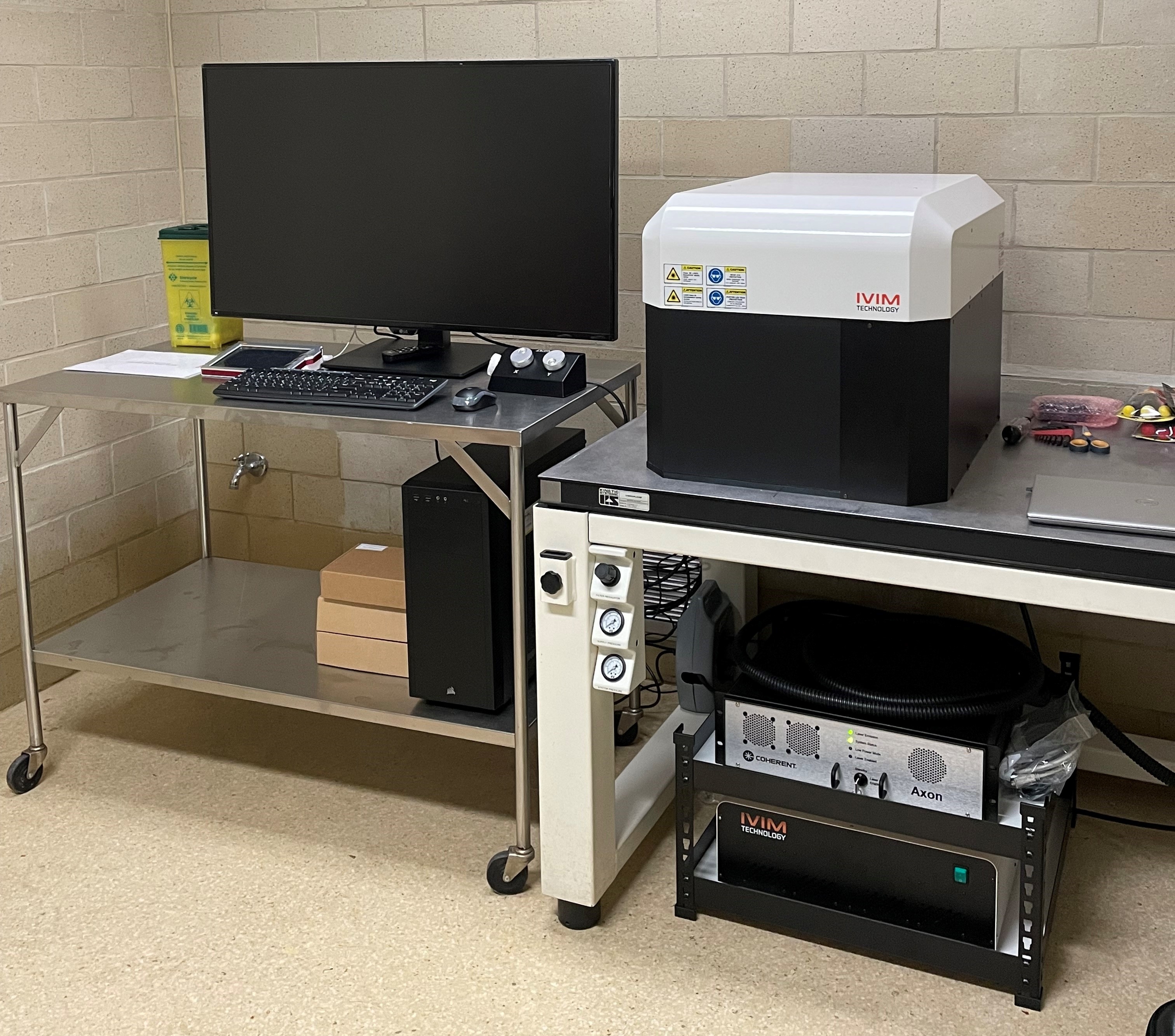
Intravital microscopy equipment
Intravital microscopy (IVM) provides imaging of numerous organs and tissues at microscopic level in live mice. This technology allows for one time as well as longitudinal imaging with capturing images and live videos. For more information, please visit the following websites:
Intravital microscope applications

Examples of images done by intravital microscopy

