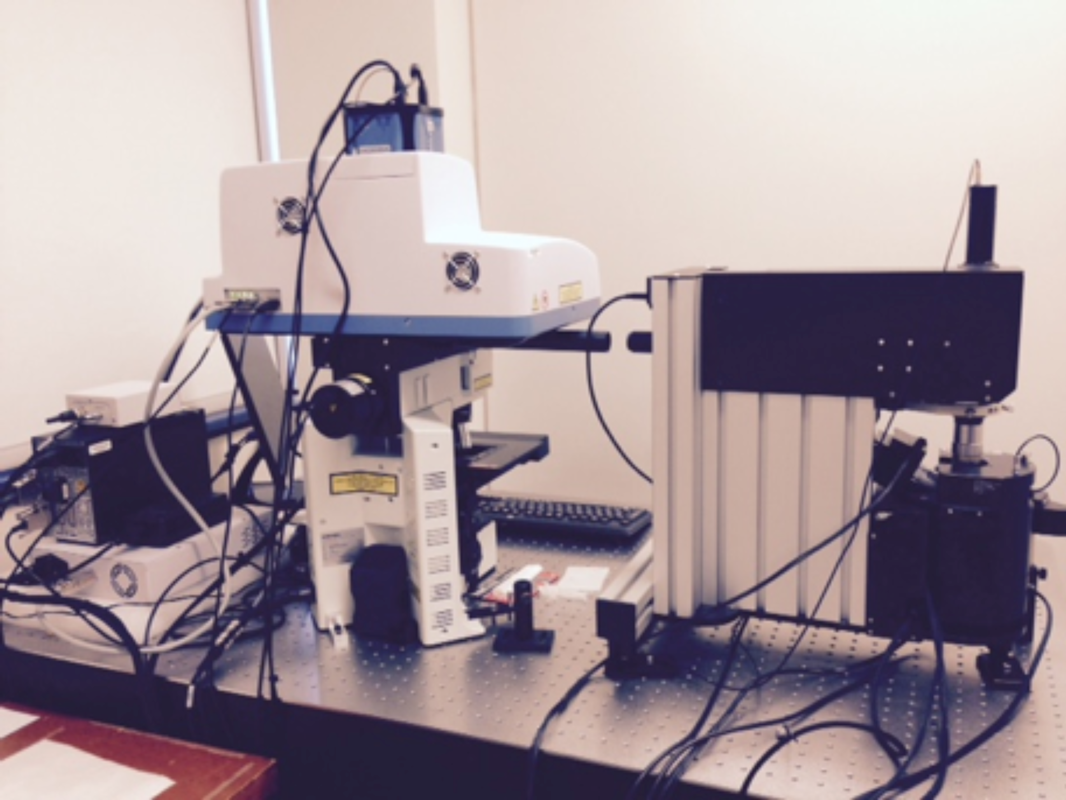
Explore the technologies available through the Microscopy Facility!

Overview

JEM-2100F FETEM (JEOL)

  • Field Emission Gun
  • Acceleration Voltage: 200kV (80, 100, 120kV)
  • TEM (0.19nm point-to point resolution)
  • HRTEM (0.1 nm lattice image)
  • STEM (0.2 nm resolution)
  • Electron Diffraction
    • Selected area electron diffraction
    • Nano-beam electron diffraction
    • Convergence beam electron diffraction
  • Energy Dispersive X-ray Spectroscopy (EDS)
    • elemental analysis and mapping
  • Electron Energy Loss Spectroscopy (EELS)
    • EFTEM
    • EELS spectrum imaging
    • EELS/EDS imaging
Machine thermostarms

FEI Tecnai G2 Spirit Twin TEM

  • Acceleration voltage: 20 – 120kV (Lab6 emitter)
  • High resolution and high contrast imaging for biological and soft materials
  • Cryo-electron microscopy
  • Electron tomography
  • Integrated Fluorescent Microscopy and Electron Microscopy (iCorr)
FEI Tecnai G2 Spirit Twin TEM

JSM-7500F FESEM (JEOL)

  • Field Emission Gun
  • Acceleration Voltage: 0-30 kV
  • Secondary Electron Imaging
    • 1.4 nm (at 1 kV)
    • 1 nm (at 15 kV))
  • Low voltage STEM
    • 1nm (at 30 kV)
  • Backscattered Electron Imaging (Z-contrast)
  • Energy Dispersive X-ray Spectroscopy (EDS)
  • Electron Backscatter Diffraction (EBSD)
Machine thermostarms

HORIBA XploRA Plus Raman Microscope

  • Two-laser  wavelengths Raman spectroscopy
  • Small area Raman analysis with Optical microscope at 10x and 100x magnifications
  • High resolution Raman imaging
  • Couples with AIST-NT OmegaScope 1000 for TERS (Tip Enhanced Raman Spectroscopy)
HORIBA XploRATM Plus Raman Microscope

AIST-NT OmegaScope 1000

  • Atomic Force Microscopy in contact mode and tapping mode
  • Kevin probe microscopy
  • Couples with HORIBA XploRa Plus Raman microscope for TERS (Tip Enhanced Raman Spectroscopy)

Sample preparation equipment

  • Precision Etching and Coating System (Gatan Model 882 PECS)
    PECS is a bench-top ion beam based etching and coating system, designed to etch and/or sputter-coat specimens for both SEM and TEM applications.
  • Ultramicrotome (Leica Ultracut UC6)
    Section thickness: 1 nm – 15 µm (with a diamond knife)
  • Turbo Pumping Station (Varian V-81M Turbo Station)
    Vacuum : 10-8 Torr (for pre-pumping TEM and SEM specimens)
  • Ultra-centrifuge (Lengend RT)
     
Rotor type Capacity Max. Speed
Swing bucket 4 x 750 ml 4150 rpm
F14-6x250LE 4 x 250 ml 10,000 rpm
F21-48x2 48 x 2 ml 15,000 rpm
  • Furnace (CARBOLITE)
    Maximum Temperature: 1700 oC
  • Vacuum oven (Lindburg-Blue M)
    Maximum Temperature: 260 oC

Fee structure

Please contact us for consultation and fees

Yun Liu
Materials Characterization Facility Manager
Telephone: (613) 562-5800, ext. 6786
[email protected]