Histological staining is used to highlight important features of the tissue as well as to differentiate structural elements of the tissue by their color and/or staining intensity. They are classified into 2 categories: Routine and Special Stains.

Histological staining

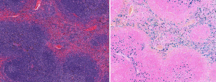
Routine Stain – Hematoxylin and Eosin (H&E)

At the Histology Core we routinely stain slides with the Hematoxylin and Eosin (H&E) stain. It provides a comprehensive picture of the microanatomy of a tissue and is frequently used by pathologists and researchers as an initial assessment.

How does H&E staining work?

It consists of 2 dyes; Hematoxylin and Eosin, which will stain the nucleus and the cytoplasm, respectively, in two different colors. Hematoxylin is a basic dye that stains the acidic components of the tissue (e.g., nucleus, ribosome, and rough endoplasmic reticulum) a purplish-blue color.  Eosin is an acidic dye that stains the basic tissue components (e.g., cytoplasm, cell walls, extracellular fibers) a reddish or bright pink color.

H&E Stain (left) vs Prussian Blue iron stain (right) in mouse spleen

H&E stain vs prussian blue iron stain

H&E Stain (left) vs Prussian Blue iron stain (right) in mouse spleen
PAS Stain (left) vs Silver Stain (right) showing basement membrane in the glomeruli

PAS stain vs silver stain

PAS Stain (left) vs Silver Stain (right) showing basement membrane in the glomeruli
H&E Stain (left) vs Alcian Blue Stain (right) showing goblet cells in the rat intestine

H&E stain vs alcian blue stain

H&E Stain (left) vs Alcian Blue Stain (right) showing goblet cells in the rat intestine

Special stains

Special stains are usually performed after H&E staining. It refers to a wide variety of alternate staining techniques and procedures used to provide more in-depth information on specific structure(s) in a sample. Rather than specific antibody binding, special staining techniques are based on simple chemical reactions such as acid-base chemistry and oxidation-reduction reactions.

Movat Pentachrome Staining in rat lung
Movat pentachrome staining in rat lung

The Histology Core Facility routinely offers the following Special Stains:

If you would like to have a stain that is not listed, please email [email protected] to enquire.

Carbohydrates

  • Periodic Acid-Schiff (PAS)
  • Periodic Acid-Schiff (PAS) diastase (Glycogen)
  • Alcian Blue (pH 0.4, 1.0, 2.5)

Connective tissue

  • Masson’s Trichrome (collagen, fibrotic tissue, cartilage)
  • Movat’s Pentachrome (collagen, elastic fibers, mucins, muscle, cartilage)
  • Verhoeff-Van Gieson (Elastic)
  • Picrosirius Red (Collagen)
  • Safranin O (Cartilage)
  • Toluidine Blue (Cartilage, Mast cells)
  • HPS (Hematoxylin Phloxine Stain: Muscle, Collagen)

Lipids

  • Oil Red O – Frozen tissue only

Microorganisms

  • Gram stain, Brown and Brent
  • Modified GMS Silver Stain (Fungi)
  • Giemsa Jenner (Parasites)

Hematology

  • Wright Giemsa (Blood Smear)

Minerals and pigments

  • Perl’s Prussian Blue (Iron )
  • Von Kossa (Calcium)
  • Alizarin Red (Calcium)
  • Congo Red (Amyloid)

Nervous system

  • Cresyl Violet (Neurons)
  • Luxol Fast Blue (LFB), LFB –PAS  (White and Grey matter)

Other stains (contact the Core if you require other stains)

  • Churukian Silver Stain (Alpha cells in pancreas, neuroendocrine cells)
  • Gomori’s Aldeyhyde Fuchsin (Beta cells in pancreas)
  • Jones Methenamine Silver ( glomerular capsule)