Immunohistochemistry (IHC) and Immunofluorescence (IF) are specialized techniques that involve the use of antibodies to detect specific proteins (antigens) in cells of a tissue section.

Immunohistochemistry

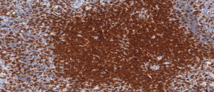
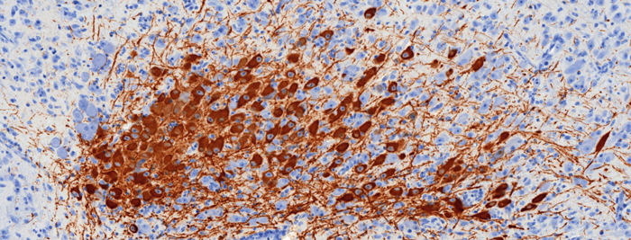
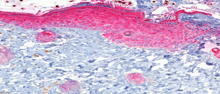
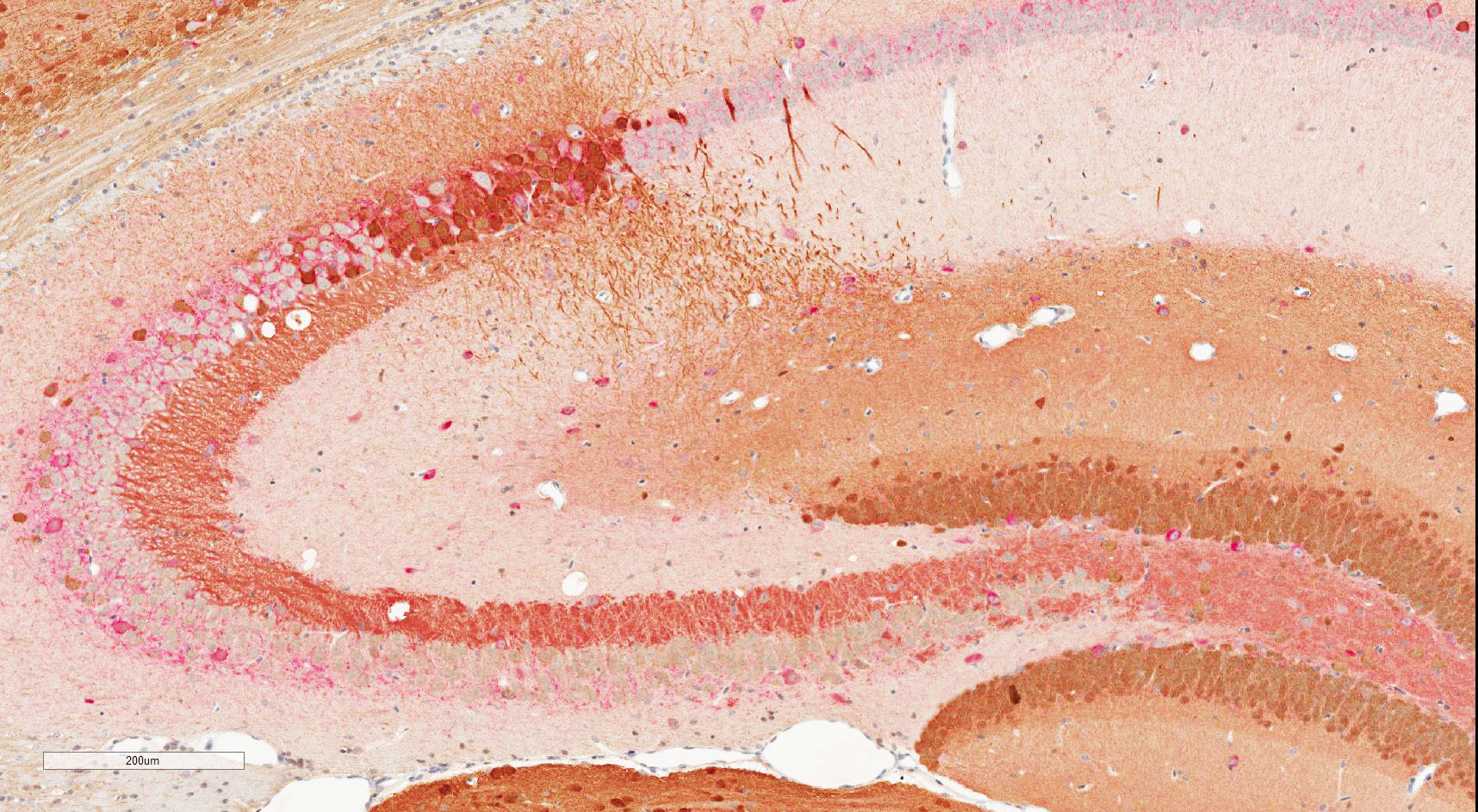
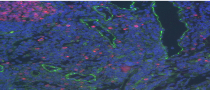
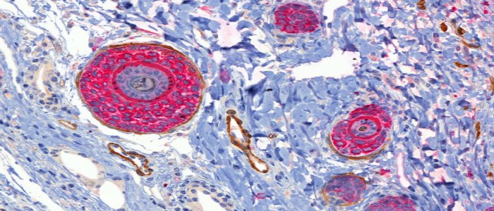
Proteins are visualized using either chromogenic detection with a colored enzyme substrate (IHC) or fluorescent detection with fluorescent dyes (IF).

Before submitting any samples for IHC or IF, we strongly encourage researchers to consult with us prior to antibody selection and assay design.

IHC (DAB) staining using CD5 on human tonsil

IHC (DAB) staining

IHC (DAB) staining using CD5 on human tonsil
IHC (DAB) staining showing dopaminergic neurons in mouse brain

IHC (DAB) staining

IHC (DAB) staining showing dopaminergic neurons in mouse brain
IHC (AP) staining using cytokeratin in mouse skin

IHC (AP) staining

IHC (AP) staining using cytokeratin in mouse skin
Double labelling by chromogenic IHC in mouse brain

Double labelling by chromogenic IHC

Double labelling by chromogenic IHC in mouse brain
Double labelling by immunofluorescence in human tonsil

Double labelling by immunofluorescence

Double labelling by immunofluorescence in human tonsil
Double labelling by chromogenic IHC in mouse skin

Double labelling by chromogenic IHC

Double labelling by chromogenic IHC in mouse skin

The Histology Core Facility offers:

  • Titration of new antibodies
  • Staining of established antibodies
  • Manual and Automated staining (Leica Bond III Immunostainer):
    • Chromogenic IHC
      1. Horseradish peroxidase (HRP) in brown
      2. Alkaline phosphatase (AP) in pink
      3. Single or dual color
    • Immunofluorescence
      1. Single or multi-color
  • In-Situ Hybridization  (Coming soon)